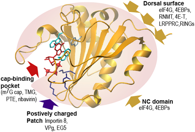
Figure 2.

The human eIF4E structure binding the m7G cap (in red) and known interactions: eIF4E structure and relative position of regions utilized by known partner proteins. The red arrow indicates the cap-binding site with the associated tryptophans in light blue, the purple arrow indicates the positively charge patch, where Importin 8, VPg and EG5 bind with the 2 lysines and the arginine shown. The dorsal surface is also shown and as is the surface used by the NC (non- canonical domain) of eIF4G and 4EBPs with gold yellow arrows (Adapted from [52], PDB3AM7).
