
Figure 1. Validation of 6-OHDA mouse model.
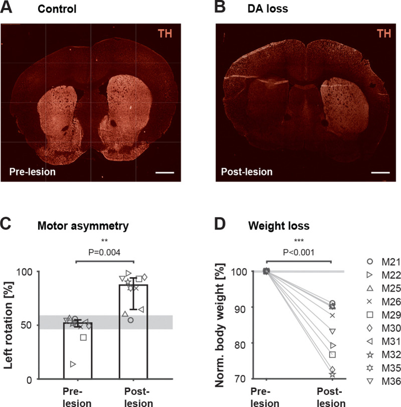
A unilateral injection of 0.2ul of 6-OHDA (15 mg/ml solution 6-OHDA.HBr) was administered into the left median forebrain bundle via Hamilton syringe. Mice were allowed to recover for up to 2 weeks before testing continued. A. Coronal sections showing the striatal hemispheres of a control mouse without lesion stained for TH expression. B. Coronal section of a 6-OHDA-lesioned mouse brain stained for TH shows over 80% reduction in fluorescence in the dopamine (DA)-depleted hemisphere compared to the control hemisphere. Across all lesioned mice, DA loss was consistently quantified via TH immunofluorescence, showing a median reduction of 77.25% (n = 8, P = 0.008, paired, two-sided Wilcoxon signed rank test). Scale bar: 1 mm. C. Percent left turns performed by each mouse in the rotameter test before and after 6-OHDA injection. Bars represent medians across mice with percentiles (n = 10). ** P = 0.004, paired, two-sided Wilcoxon signed rank test. D. Body weight before and after 6-OHDA injection. Percent body weight is calculated by identifying the minimum weight within the recovery period. Numbers are normalized to each animals’ weight on the day before the lesion (n = 10). *** P < 0.001, paired, two-sided Wilcoxon signed rank test. Gray areas represent median range across sham operated mice (n = 2).