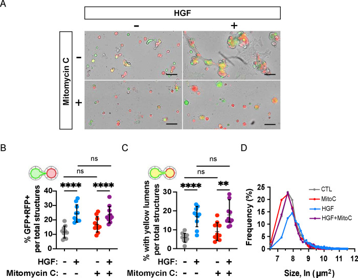
Figure 4. Proliferation is dispensable for HGF-induced anastomosis.
Replated spheroids were treated with either HGF (10 ng/mL), an inhibitor of cell proliferation, mitomycin C (2.5 μM), or a combination, for two days. (A) Representative images of replated spheroids after treatment period. Scale bars = 200 μm. (B) Percentage of GFP+-RFP+ structures were not altered by inhibitor treatment. (C) Percentage of yellow lumens was unaffected by mitomycin C. (D) Cross-sectional area of structures at endpoint. Data shown are representative of at least four independent experiments performed in triplicate. Measures of interconnection were compared by two-way ANOVA, P<.0001****.

