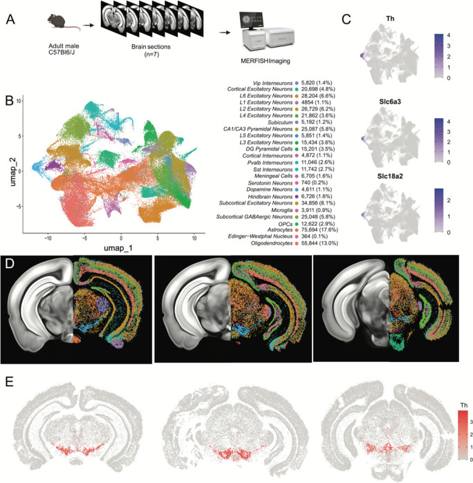
Figure 3.

Identification of dopamine neurons in MERFISH. A) Schematic showing the processing and imaging of brain tissue with MERFISH. B) Clustering and annotation of all cells identified by MERFISH. C) Relative expression of Th, Slc6a3 and Slc18a2 shows the presence of a single cluster (blue cluster in B) that expresses all three genes. D) Spatial location of neuronal clusters for whole brain MERFISH along three rostral-caudal delineations associated with −3.0, −3.2 and −3.6mm Bregma. E) Cellular expression of Th in the sections shown in D.
