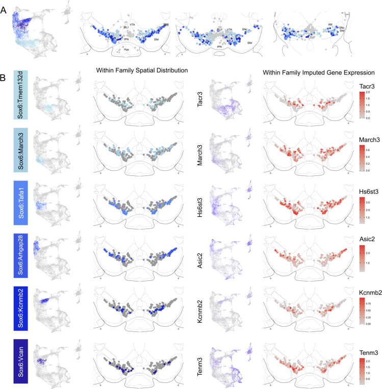
Figure 5.
Spatial localization of Sox6 family of DA neurons. A) Distribution of Sox6 family DA neuron subtypes relative to all other DA neurons along the rostral-caudal axis. DA cells belonging to the Calb1 and Gad2 family are colored in light grey. B) Location of individual subtype within the Sox6 family (left two panels) with other cells in the Sox6 family shown in dark grey. Relative cellular expression of genetic markers associated with each subfamily shown in a UMAP and a midbrain section.

