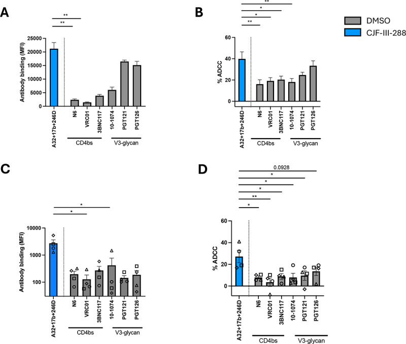
Figure 4. The A32/17b/246D/CJF-III-288 cocktail mediates ADCC more efficiently than bNAbs.

(A) HIV-1CH058TF-infected primary CD4+ T cells were stained with 5 μg/mL of total antibodies in presence of either DMSO depicted in gray or CJF-III-288 depicted in blue 48hrs post-infection. Flow cytometry was performed to detect antibody binding. The graph represents the MFI of Alexa-Fluor 647. (B) HIV-1CH058TF-infected primary CD4+ T cells were incubated with 5 μg/mL of total antibodies in presence of either DMSO depicted in gray or CJF-III-288 depicted in blue. CD4+ T cells were used as target cells, while autologous non-infected PBMCs were used as effector cells in our FACS-based ADCC assay. The graph represents the percentage of ADCC obtained in presence of the indicated antibodies in at least 4 independent experiments. (C) Cell-surface staining of primary CD4+ T cells isolated from 4 HIV-1-infected individuals under ART after ex-vivo expansion with 5 μg/mL of total antibodies in presence of either DMSO depicted in gray or CJF-III-288 depicted in blue. Each symbol represents a different donor. Flow cytometry was performed to detect antibody binding. The graph represents the MFI of Alexa-Fluor 647. (D) ADCC was assessed on primary CD4+ T cells isolated from 4 HIV-1-infected individuals under ART after ex-vivo expansion. CD4+ T cells were used as target cells, while autologous PBMCs were used as effector cells in our FACS-based ADCC assay with 5 μg/mL of total antibodies in presence of either DMSO depicted in gray or CJF-III-288 depicted in blue. The graph represents the percentage of ADCC obtained in presence of indicated antibodies. Statistical significance was tested using (A, C) Kruskal- Wallis (B, D) One-way ANOVA, according to population normality (*, P < 0.05; **, P < 0.01).
