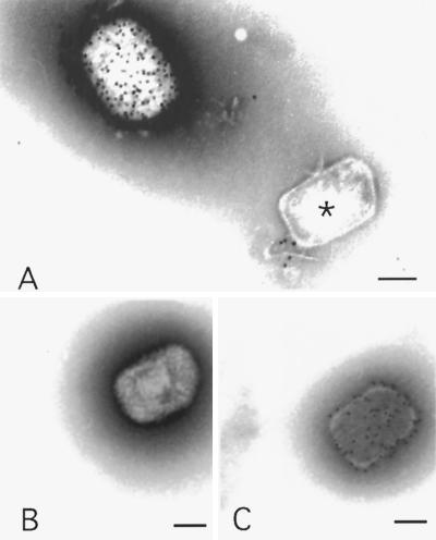
FIG. 3.
Negative staining EM and immunolabeling of intact IMVs and actD cores. (A) Labeling with anti-p14. The upper left shows a heavily labeled IMV; in the lower right an unlabeled core is evident. (B and C) labeling with anticore, showing an unlabeled IMV (B) and a labeled actD core (C). Bars = 100 nm.

