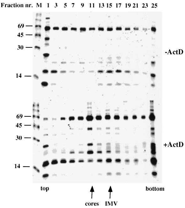
FIG. 4.
35S-labeled pattern of subviral components isolated from VV-infected cells separated on sucrose gradients. Gradient fractions from Fig. 1B were separated by SDS-PAGE on a 15% gel, which was then processed for autoradiography. The upper panel represents gradient fractions from untreated (-ActD) cells, while the lower panel is from treated (+ActD) cells. The top and bottom of the gradients, as well as positions of the core and IMV peak, are indicated. In each case, fractions with even and odd numbers were pooled such that, e.g., fraction 1 denotes the combined radioactive pattern of fractions 1 and 2. M, 14C-labeled markers of 14, 30, 45, and 69 kDa.

