Figure 5.

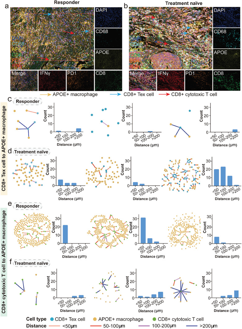
Spatial mIHC staining distinguishes the distance between APOE+ macrophages and CD8+ Tex cells and CD8+ cytotoxic T cells in human TNBC tissues. a,b) The mIHC images of indicated APOE+ macrophages (orange arrows), CD8+ Tex cells (blue arrows), and CD8+ cytotoxic T cells (red arrows) in ICI responder (left) or TN (right) TNBC tumor sections. c,d) The distances and their measurement between APOE+ macrophages and CD8+ Tex cells in ICI responder and TN patients. e,f) The distances and its measurement between APOE+ macrophages and CD8+ cytotoxic T cells in ICI responder and TN patients. mIHC: multiplex immunohistochemistry; ICI: immune checkpoint inhibitor; TN: treatment naïve; TNBC: triple negative breast cancer.
