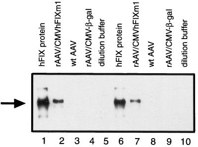
FIG. 3.
Western blot analysis of rAAV virion-packaged proteins. rAAV preparations diluted to 1011 particles/ml were resolved on an SDS–10% polyacrylamide gel and electroblotted onto polyvinylidene difluoride membranes. The hFIX proteins on blots were visualized by the chemiluminescent detection system. Lanes 2 to 4 and 7 to 9 were loaded with 4 or 2 μl of rAAV vectors, respectively. Lanes 1 and 6 were loaded with 8 and 4 ng of purified plasma-derived hFIX protein, respectively. Lanes 2 and 7 were loaded with rAAV/CMVhFIXm1. Lanes 3 and 8 and lanes 4 and 9 were loaded with wild-type AAV or rAAV/CMV-β-gal, respectively. Lanes 5 and 10 contain the dilution buffer alone. The arrow indicates the position of the hFIX protein (56 kDa).

