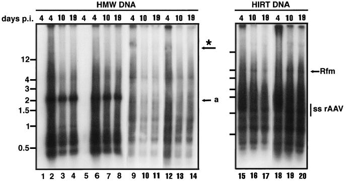
FIG. 4.
Southern blot analysis of DNA isolated from rAAV/CMVhFIXm1-transduced cells. HMW and Hirt DNA were isolated from muscle cells transduced at a MOI of 105 and maintained in F10-based medium (Fig. 2I and J). HMW or Hirt DNA was isolated 4 days p.i. (lanes 2, 6, 9, 12, 15, and 18), 10 days p.i. (lanes 3, 7, 10, 13, 16, and 19), or 19 days p.i. (lanes 4, 8, 11, 14, 17, and 20). Lanes: 2 to 4 and 9 to 11, HMW DNA from cells transduced at the myoblast stage; 6 to 8 and 12 to 14, HMW DNA from cells transduced at the myotube stage; 1 and 5 respective mock-infected control DNA isolated on day 4. HMW DNA (4 μg) was digested with EcoRI and subjected to Southern blot analysis with the CMV promoter probe. Lanes 1 to 8 contain EcoRI-digested HMW DNAs, and lanes 9 to 14 contain undigested HMW DNAs. Arrow a indicates the position of the 1.9-kb internal EcoRI fragment. The possible position for the >12-kb genomic DNA signal, more prominant in Fig. 5, is shown by an asterisk. Lanes 15 to 17 contain Hirt DNA from cells transduced at the myoblast stage; lanes 18 to 20 contain Hirt DNA from cells transduced at the myotube stage. Hirt DNA isolated from cells cultured in the rich medium tends to give less discernible bands, and undigested or digested genomic DNA show bands of >4-kb (lanes 9 to 14) and <1.9 kb (lanes 2 to 4 and 6 to 8), respectively. These bands, however, are not seen when genomic DNA prepared from rAAV-transduced cells was grown in differentiation medium (see Fig. 5). At present, little is known about why genomic DNA from cells maintained in rich medium with much active metabolism tends to have these bands.

