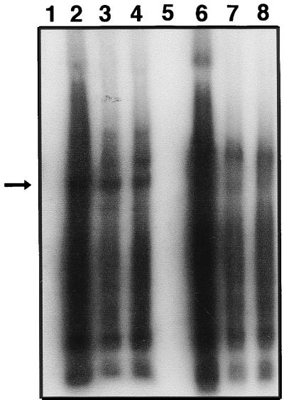
FIG. 6.
Detection of head-to-tail junctions in HMW DNA. HMW DNA (4 μg) isolated from transduced myotubes (Fig. 2I) were digested with BstBI (lanes 1 to 4) or NotI (lanes 5 to 8) and analyzed by Southern blot analysis with the CMV promoter-specific probe. HMW DNA was isolated 4 days p.i. (lanes 2 and 6), 10 days p.i. (lanes 3 and 7), or 19 days p.i. (lanes 4 and 8). Lanes 1 and 5 contain DNA from mock-infected controls. BstBI recognizes a single site in the vector. The arrow indicates the expected head-to-tail junction. No NotI site exists in the vector. NotI digestion did not release any distinct band and showed a profile similar to that of undigested samples (Fig. 4, lanes 9 to 14).

