. 2024 May 31;26(24):16980–16988. doi: 10.1039/d4cp01268b

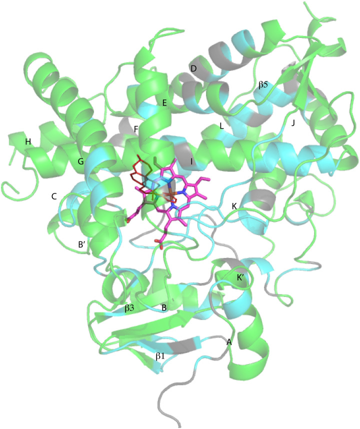
Fig. 3. CYP17A1 (PDB entry 3RUK) with abiraterone bound, showing distribution of sequential backbone resonance assignments. Assignments for regions in green are supported by multiple forms of data (see text for details), regions in blue are provisionally assigned based on fewer data. Those shown in grey are not assigned. Secondary structures were assigned by DSSP analysis of PDB 3RUK (see ESI) are labeled as first described by Poulos.23 Helices: A, M49–Y60; B, H80–L86; B′, A100–A105; C, A119–A133; D, L142–T159; E, S168–F184; F, E194–L209; G′, W120–I123; G, T228–K253; H, M262–N272; I, D289–H320; J, P322–N335; K, L351–L363; K′, L396–H401; K′′, P414–F417; L, E445–R462. Strands: β1, I63–M68, K71–V76, E391–I394, H373–K374; β2, S379–I381, F384–386; β3, F463–E466, K492–V495; β4, I479–P480, F484–L385; β5, F463–Q466, V491–495. Abiraterone is shown in thin red lines, heme is shown in heavier pink lines.

Fig. 3