. 2024 May 31;26(24):16980–16988. doi: 10.1039/d4cp01268b

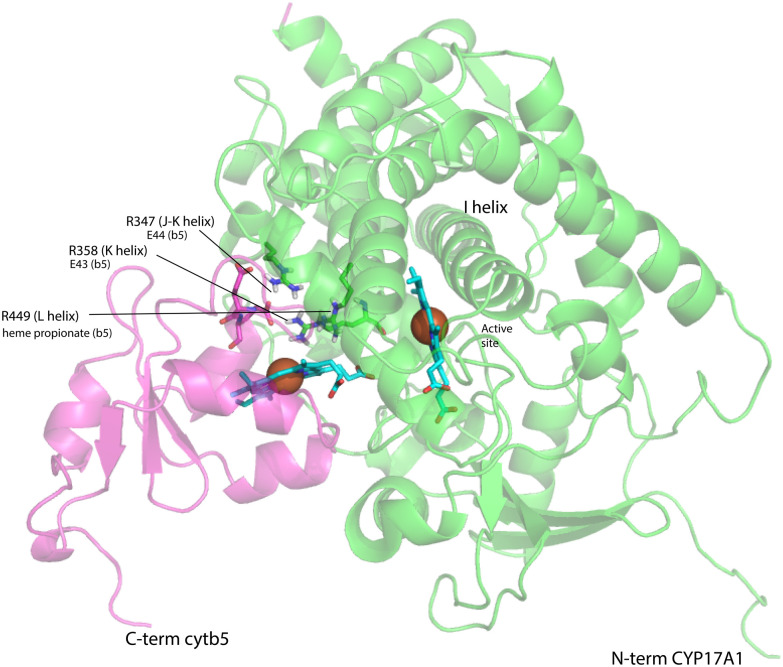
Fig. 9. Model for a complex formed by cytb5 on the proximal face of CYP17A1, placing Arg 347, Arg 358 and Arg 449 adjacent to Glu 44, Glu 43 and a cytb5 heme propionate, respectively. Both heme iron atoms are shown as orange spheres. CYP17A1 is rotated counterclockwise ∼90° and tilted clockwise ∼20° from the orientation in Fig. 3 membrane binding domains of both proteins are not shown, but are extensions of the C-term of cytb5 and N-term of CYP17A1 that are oriented toward the heavy black line at the bottom of the figure. Crystal structures 1ICC and 3RUK were used as templates. Model is courtesy of Prof. Eliana Asciutto, UNSAM, Buenos Aires, AG.

Fig. 9