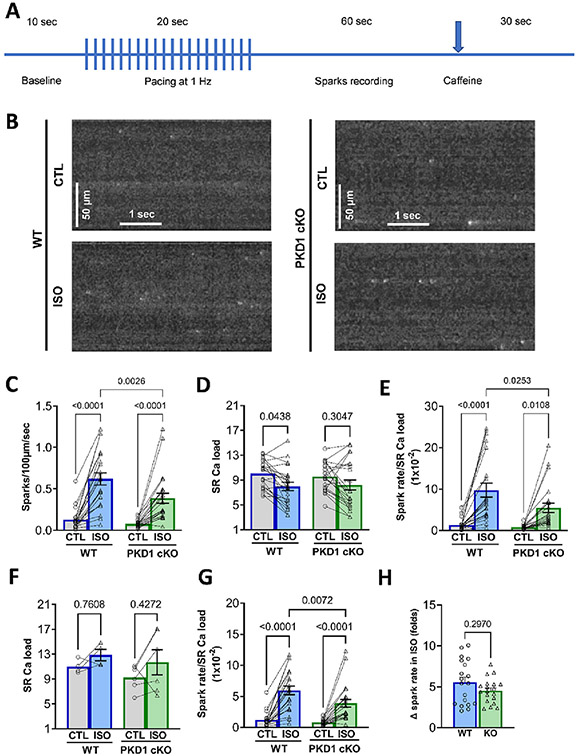
Fig. 2.
ISO-induced increase in spark rate is blunted in PKD1 cKO ventricular myocytes. A, Experimental protocol for Ca2+ spark measurements. B, Representative confocal images of diastolic Ca2+ sparks in both WT and PKD1 cKO myocytes at baseline and after 5-min incubation with 100 nM isoproterenol (ISO). Linescan image contrast was enhanced using Matlab™ software to improve visibility of Ca sparks. C, Diastolic Ca2+ sparks rate significantly increased more in WT myocytes. D, ISO-induced SR Ca2+ load decrease is similar in WT and PKD1 cKO ventricular myocytes. E, Normalization of spark rate by SR Ca2+ load in WT and PKD1 cKO. F, SR Ca2+ load measured immediately after pacing. G, Normalization of spark rate by SR Ca2+ load in panel F. H, Fold-change in response to ISO for Ca2+ spark frequency in WT and PKD1 cKO. Data points represent cells (WT, n = 22; KO, n = 23). Mice: WT, N = 11; KO, N = 12. Data are presented as mean ± SEM. Two-way ANOVA, followed by Tukey’s multiple comparisons test. Differences were considered statistically significant if P < 0.05.

