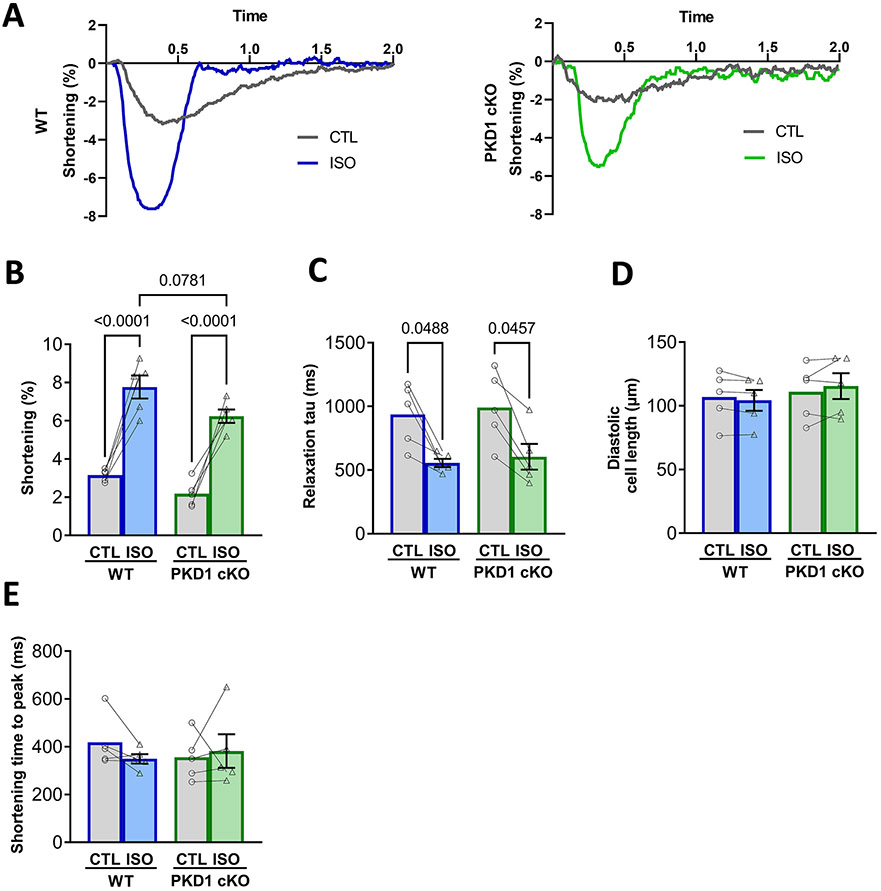
Fig. 4.
Both WT and PKD1 cKO ventricular myocytes increase contractility similarly after β–AR stimulation (isoproterenol). A, Representative traces of cell shortening in WT and PKD1 cKO myocytes. B, Fractional shortening amplitude, relaxation tau decay, time to peak of contraction and diastolic cell length were similar in both WT and PKD1 cKO myocytes at baseline and after 5-min incubation with 100 nM isoproterenol (ISO). Data points represent cells (WT, n = 6; KO, n = 6). Mice: WT, N = 4; KO, N = 4. Data are presented as mean ± SEM. Two-way ANOVA, followed by Tukey’s multiple comparisons test. Differences were considered statistically significant if P < 0.05.

