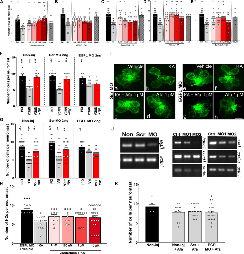
Fig. 3. EGFR inhibitors protect against hair cell excitotoxicity in zebrafish.
(A to E) Five- to 6-dfp zebrafish were incubated with 500 μM of KA for 1 hour followed by 2-hour incubation with different concentrations of EGFR inhibitors. (A) Osimertinib, (B) JCN037, (C) dacomitinib, (D) afatinib, and (E) zorifertinib. *P < 0.05, **P < 0.01, and ***P < 0.001 versus KA alone. (F to K) Zebrafish non-injected or injected with 2 ng of scrambled or EGFL-specific morpholinos. Animals (3 dpf) were preincubated with 500 μM of NMDA (F) or KA (G) for 1 hour followed by a 2-hour incubation with vehicle or afatinib (1 μM). Quantification of the HCs was performed in SO3, O1, and O2 neuromasts. **P < 0.01 and ***P < 0.001 versus ototoxin alone. ɸP < 0.05, ɸɸP < 0.01, and ɸɸɸP < 0.001 versus the scrambled morpholino. (H) Zebrafish EGFL knockdowns (KDs) incubated with KA followed by different concentrations of zorifertinib. ****P < 0.01 versus KA alone. (I) Representative images of scrambled and EGFL morphants with the different treatments. Green (GFP) denotes the neuromast hair cells. Ctrl, control; KA + Afa 1 mM, KA and afatinib; Afa 1 mM, afatinib only. (J) Left gels: Confirmation of EGFL KD by reverse transcription polymerase chain reaction of scrambled and EGFL morphants. Right gels: EGFR downstream effectors after EGFL KD. Ctrl, control; MO1, EGFL MO 2 ng; MO2, EFGL MO 4 ng. hdac1, ccnd1, aurka, cox1, and cox2a. (K) Hair cell quantification in the different morphants under baseline conditions. Hair cell quantification is expressed as means ± SEM. N = 5 to 6 for each group. One-way ANOVA for (A) to (H) and (K).

