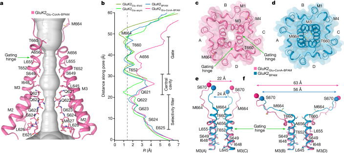
Fig. 4. Open pore in GluK2Glu–ConA–BPAM.
a, Pore-forming segments M2 and M3 in GluK2Glu–ConA–BPAM, with residues lining the pore shown as sticks. Only two subunits (A and C) of the four are shown, with the front and back subunits (B and D) omitted for clarity. The pore profile is shown as a space-filling model (grey). The green arrow indicates the gating hinge where M3 bends during channel opening. b, Pore radius for GluK2BPAM (blue), GluK2Glu–ConA–BPAM (pink), GluK2Glu-4fold (dirty green) and GluK2Glu-asym (green) calculated using HOLE. The vertical dashed line denotes the radius of a water molecule (1.4 Å). c,d, Extracellular view of the ion channel semi-transparent surface in GluK2Glu–ConA–BPAM (c) and GluK2BPAM (d; PDB ID: 8FWS). e,f, Superposition of the pore-lining segments M3 and M3–S2 linkers in subunits A and C (e) and B and D (f), with residues lining the pore in the gate region shown as sticks. Distances between Cα atoms of S670 are indicated.

