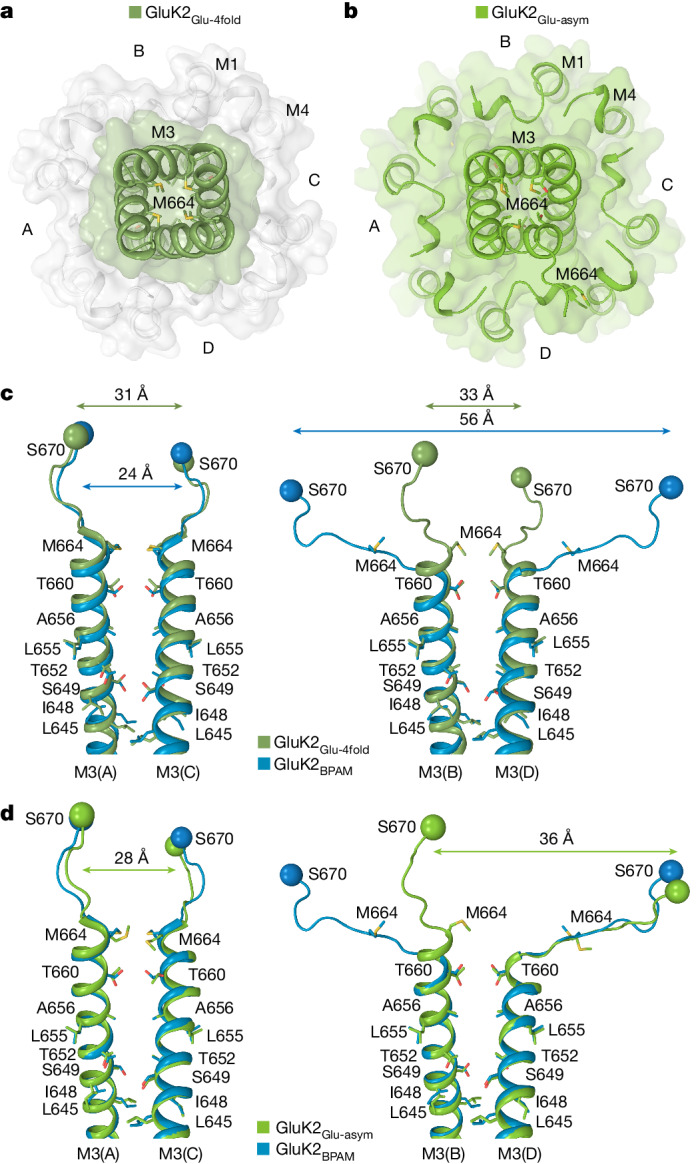
Fig. 6. Closed pore of GluK2Glu-4fold and GluK2Glu-asym.

a,b, Extracellular view of the ion channel semi-transparent surface in GluK2Glu-4fold (a) and GluK2Glu-asym (b). c,d, Superposition of the pore-lining segments M3 and M3–S2 linkers in subunits A and C (left) and B and D (right) of GluK2BPAM (PDB ID: 8FWS, blue) and GluK2Glu-4fold (c; dirty green) or GluK2Glu-asym (d; green), with residues lining the pore in the gate region shown as sticks. Distances between Cα atoms of S670 are indicated.
