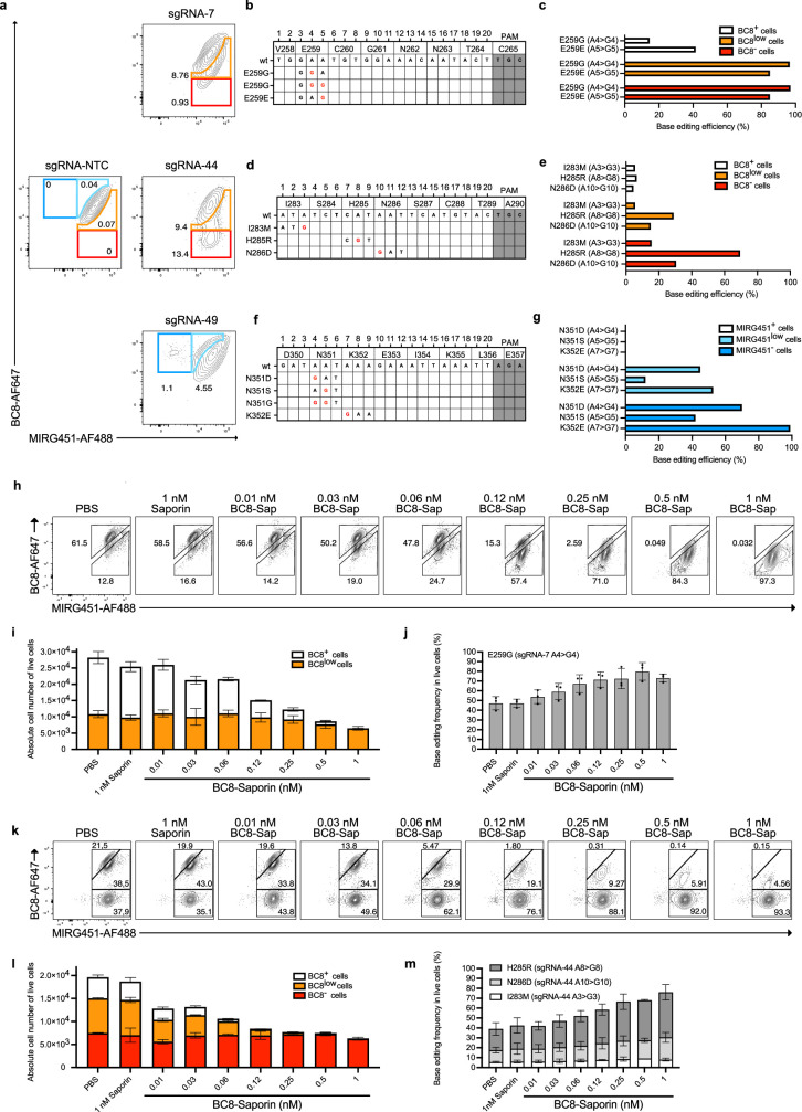
Extended Data Fig. 3. Base editing shields primary cells from a CD45 surrogate ADC.
a, Flow cytometry of human activated T cells 5 days after electroporation of ABE8e-NG mRNA with sgRNA-NTC, sgRNA-7, sgRNA-44 or sgRNA-49. b, sgRNA-7 mapped on corresponding gDNA codons c, Bar graphs of Sanger sequencing results showing amino acid substitutions in sorted populations from sgRNA-7 edited cells (matching colour code). d, sgRNA-44 mapped on corresponding gDNA codons e, Bar graphs of Sanger sequencing results showing amino acid substitutions in sorted populations from sgRNA-44 edited cells (matching colour code). f, sgRNA-49 mapped on corresponding gDNA codons g, Bar graphs of Sanger sequencing results showing amino acid substitutions in sorted populations from sgRNA-49 edited cells (matching colour code) (In (c,e,g), n = 2 biological replicates). h, Flow cytometry panels of base edited human activated T cells (ABE8e-NG + sgRNA-7) incubated with increasing concentration of BC8-Saporin for 3 days. i, Quantification of the absolute number of living cells post-killing from (h) and their BC8 phenotype. j, Bar graphs showing the percentage of E259G (sgRNA-7 A4 > G4) base conversion in live cells for each BC8-Saporin concentration. k, Flow cytometry panels of base edited human activated T cells (ABE8e-NG + sgRNA-44) incubated with increasing concentration of BC8-Saporin for 3 days. l, Quantification of the absolute number of living cells post-killing from (k) and their BC8 phenotype. m, Bar graphs showing the percentage of E259G (sgRNA-7 A4 > G4) base conversion in live cells for each BC8-Saporin concentration (In (i,j,l,m), n = 3 biological replicate containing each 3 technical replicates). Each bar graph from c, e, g, i, j, l and m is plotting mean values +/− SD of biological replicates.

