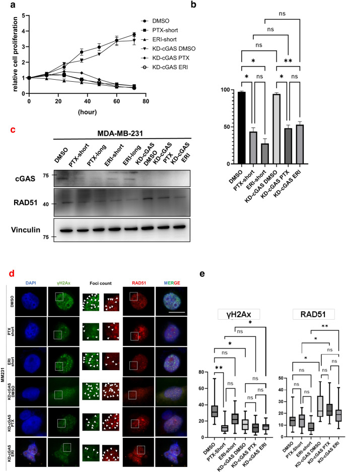
Figure 3.
The effects of KD-cGAS on cell proliferation was evaluated. MM231 cells (DMSO, PTX-short, ERI-short, KD-cGAS-DMSO, KD-cGAS-PTX, KD-cGAS-ERI) were used (a) Proliferation assay was performed. 24 h after knockdown of cGAS was defined as 0 h, and cell proliferation was evaluated every 12 h. (b) Tripan blue stain was used to evaluate the percentage of live and dead cells 24 h after treatment. (c) We evaluated the effects of knocking down of cGAS on RAD51 expression. Vinculin was used as loading control. (d) Immunofluorescence with RAD51 and γH2Ax was performed. DAPI was used for nuclear staining. The white line is 10 μm. (e) We counted the number of nuclear foci in each cell stained with γH2Ax and RAD51 on average. The meaning of the asterisks are as follows: *p < 0.05, **p < 0.01.

