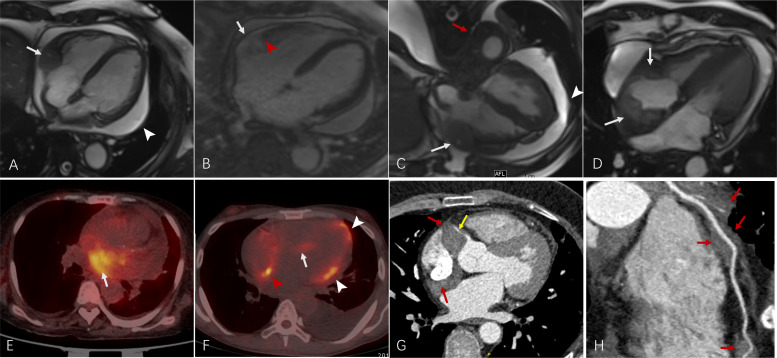
Figure 3.
(A–D) Four-chamber cardiac magnetic resonance cine images findings of patients with Erdheim-Chester disease (ECD) with cardiac involvement. (A) Right atrioventricular sulcus pseudomass surrounding right coronary artery (white arrow) and pericardial effusion (white arrowhead). (B) Right atrioventricular sulcus pseudomass (white arrow) and adjacent right ventricular myocardium (red arrowhead) enhancing on enhancement sequence. (C) Right atrioventricular sulcus pseudomass surrounding right coronary artery (white arrow) and pericardial effusion and pericardial thickening (white arrowhead) and coated aorta (red arrow). (D) Right atrium pseudomass (white arrow). (E–F) Positron emission tomography/CT fusion of patients with ECD with cardiac involvement. (E) Left atrium involvement showed 18F-fluorodeoxyglucose (FDG) uptake (white arrow). (F) Right atrium (red arrowhead), interventricular septum (white arrow) and right ventricle (white arrowhead) involvement showed FDG uptake. (G–H) Coronary artery enveloping and stenosis from right atrioventricular sulcus infiltration on CT scan. (G) The pseudomass at the right atrioventricular sulcus (red arrow) encases the origin of the right coronary artery (RCA), leading to RCA stenosis (yellow arrow). (H) The infiltration surrounds the left anterior descending artery without causing narrowing at this level.

