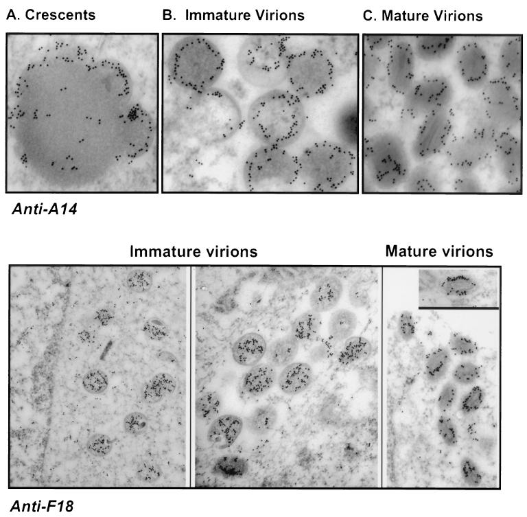
FIG. 4.
Immunoelectron microscopic localization of the A14 and F18 proteins within wt vaccinia virions. Cells were infected with wt virus (MOI, 2); at 16 hpi, cells were fixed in situ and processed as described in Materials and Methods. Sections were labeled with anti-A14 or anti-F18 serum and a secondary serum conjugated to 10-nm-diameter gold particles. (A to C) A14 is immunolocalized within the membranes of emerging crescents, immature virions, and mature virions. Magnifications, ×59,000 (A), ×56,000 (B), and ×60,000 (C). (Lower panels) F18 is immunolocalized within the interiors of immature virions. Within mature virions, it appears to localize in a ring that outlines the periphery of the lucent core. Magnifications, ×23,000 (left), ×35,000 (middle), and ×34,000 (right).

