
FIGURE 1.
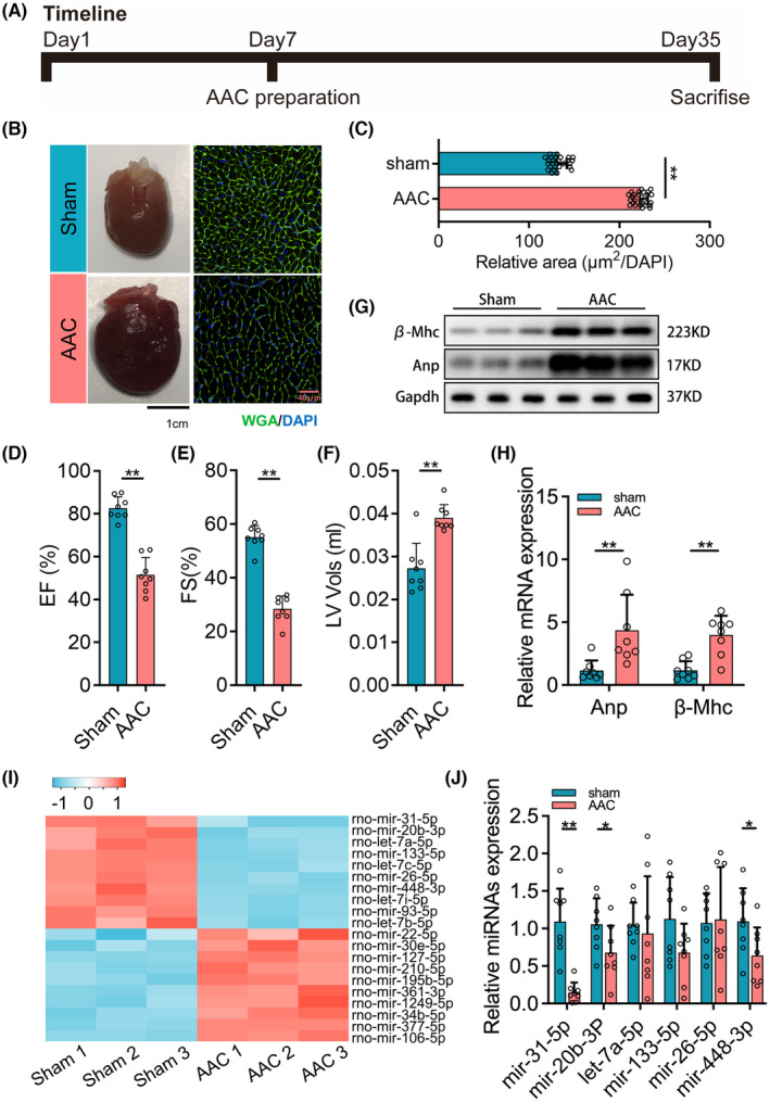
miR‐31‐5p is downregulated in abdominal aorta coarctation (AAC)‐induced myocardial hypertrophy and dysfunction. (A) The timeline of rat AAC experiments (n = 8). (B) The representative pictures of the heart and wheat germ agglutinin (WGA) staining of the sham group and AAC group. (C) The relative area of the myocardial cell surface (n = 8, 3 fields per sample). (D–F) Echocardiography analyses of cardiac function of sham and AAC groups (n = 8). (G, H) The expression of myocardial hypertrophy biomarkers including Anp and β‐Mhc was measured by western blot (n = 6) and qPCR (n = 8, 3 repeats). (I) Heat map for the top 10 upregulated and downregulated microRNAs in the AAC‐induced group compared with the sham group (n = 3). (J) The verification for the altered microRNAs presented in the heat map (n = 8, 3 repeats). For two groups, unpaired t‐tests were used. And for comparison of more than two groups, one‐way ANOVA analysis was processed, and the significance is expressed as follows: *p < 0.05, **p < 0.01.