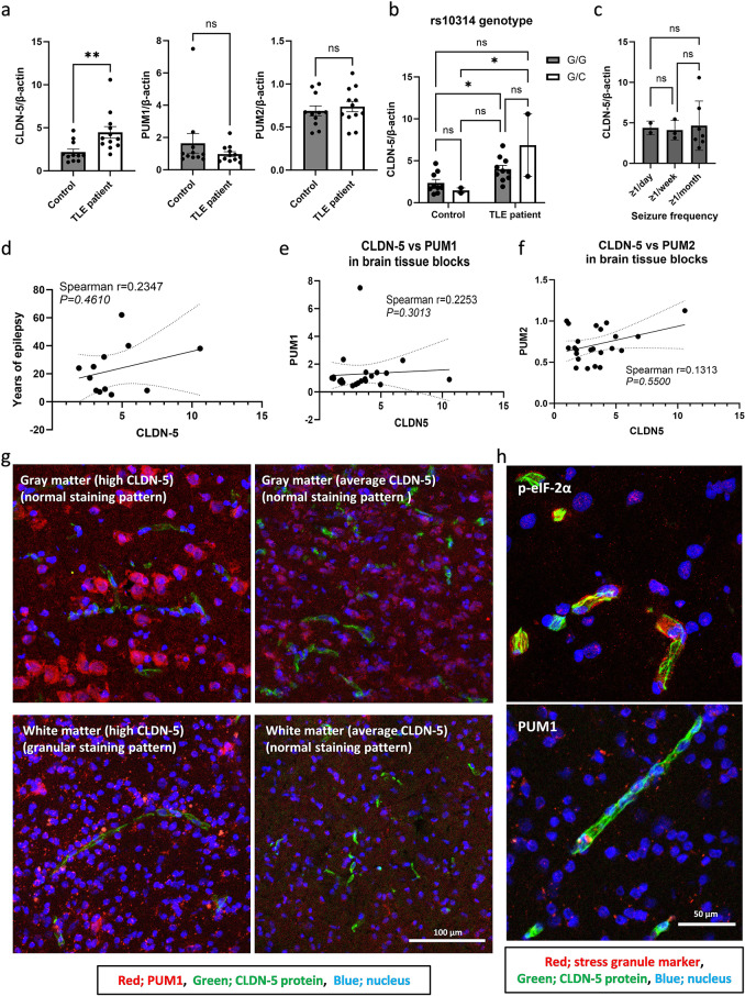
Fig. 5.
Histological localization of PUM1 in brain tissue from patients with epilepsy. (a to f) The expression level of CLDN-5 mRNA in resected human brain samples. The RNAs were isolated from the resected brain tissues obtained from control subjects (n = 11) and patients with epilepsy (n = 12). (a) CLDN-5, PUM1 and PUM2 mRNA expression levels between samples collected from control and epilepsy patients. CLDN-5 mRNA levels in the samples were broken down by (b) the difference of rs10314 genotypes (G is wild-type allele and C is rs10314 allele) and (c) seizure frequency. The data represents mean ± SD. ns, not significant (p > 0.05); *p value ≤ 0.05; **p value ≤ 0.01. The correlation between CLDN-5 mRNA and (d) years of epilepsy, (e) PUM1 and (f) PUM2 mRNA levels. Spearman r values and P values are indicated. (g) Representative confocal images of human brain sections stained with anti-PUM1 antibody. A part of the hippocampal region showed a granular PUM1 staining pattern. Red; PUM1, Green; CLDN-5. Blue; nuclei. (h) The evaluation of the induction of stress granules in vascular endothelial cells in human brain sections (high CLDN-5). Sections were stained with anti-CLDN-5 (green) and stress granule marker, anti-p-eIF-2α or anti-PUM1 (red). Nuclei (blue) were counterstained with Hoechst

