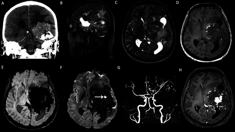
Fig. 1.

A left cerebral hemisphere giant cavernous malformation (CM) in a 33-year-old male patient with seizure and headache. (A) Coronal computed tomography (CT) scan demonstrates a large heterogeneous calcified mass in the left cerebral hemisphere. (B) Sagittal T2-weighted image (T2WI), (C) axial T2WI and (D) axial T1WI show that the mass demonstrates heterogeneous signals with “salt and pepper” appearance on nonenhanced T1WI. (E ) Susceptibility-weighted imaging (SWI) demonstrates intense “blooming artifacts” due to calcifications and chronic hemorrhage. (F) The mass is mainly hypointense on diffusion-weighted imaging (DWI) and includes a focal-restricted diffusion due to a recent hemorrhage ( arrow ). (G) Axial view of the maximum intensity projection (MIP) of the time-of-flight magnetic resonance (MR) angiography shows the cavernoma vessels in the left cerebral hemisphere. (H) Contrast-enhanced T1WI demonstrates heterogeneous enhancement.
