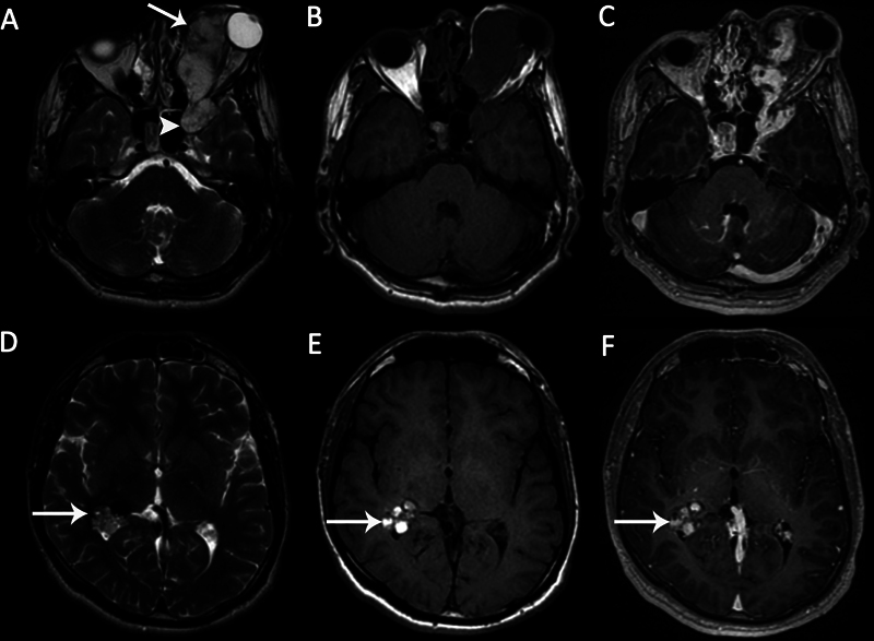
Fig. 10.

Large cavernous malformation in a 20-year-old male patient with painless proptosis of the left eye. (A) Axial T2-weighted image (T2WI) demonstrates a left orbital lobulated heterogeneous hyperintense lesion ( arrow ), extending to the anterior part of the left cavernous sinus ( arrowhead ). (B) The mass is hypointense on axial T1-weighted image (T1WI). (C) The mass shows early nonhomogeneous intense enhancement. Axial (D) T2WI and (E) axial T1WI demonstrate an incidental intraventricular cavernoma with cystic components in the right trigon ( arrows ). There are hemorrhagic signals within the lesion. (F) There is no enhancement on axial contrast-enhanced T1WI ( arrow ).
