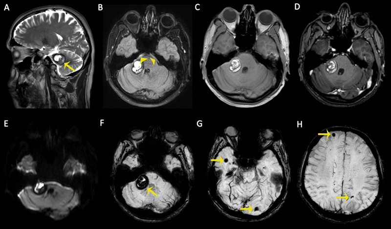
Fig. 7.

Magnetic resonance (MR) images of a 42-year-old male patient with an acute severe headache. (A) Sagittal T2-weighted image (T2WI) demonstrates a cavernous malformation (CM) in the cerebellopontine angle with peripheral hypointense hemosiderin ring ( arrow ). (B) Axial fluid-attenuated inversion recovery (FLAIR) sequence demonstrates a CM and perimesencephalic subarachnoid hemorrhage ( arrowheads ). (C) The lesion is heterogeneously hyperintense on nonenhanced T1-weighted image (T1WI) due to intralesional recent hemorrhage. (D) No enhancement was observed on axial contrast-enhanced T1WI. (E) Recent hemorrhages within the CM cause diffusion restriction on diffusion-weighted imaging (DWI) as hyperintense signals. (F–H) Susceptibility-weighted images (SWIs) show multiple intracranial CMS with “blooming artifacts” ( arrows ).
