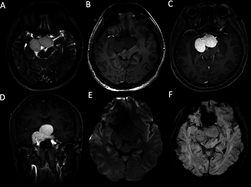
Fig. 9.

A right cavernous sinus cavernous hemangioma extending to the intrasellar and suprasellar areas in a 37-year-old male patient with a history of progressive headache and blurring of vision. (A) Axial T2-weighted image (T2WI) demonstrates a large lobulated shape mass with a homogeneous hyperintense signal compressing the optic chiasm. The mass (B) is hypointense on axial T1-weighted image (T1WI) and (C) shows homogeneous intense enhancement on contrast-enhanced axial T1WI and (D) contrast-enhanced coronal T1WI. (E) No diffusion abnormality is detected on diffusion-weighted imaging (DWI). (F) There is no “blooming artifact” on susceptibility-weighted imaging (SWI).
