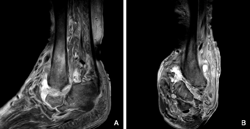
Figure 2.

Sagittal (A) and coronal (B) T2 magnetic resonance imaging show bone resorption of the talus, heel, cuneiform and navicular bone accompanying with edema of the bone marrow of distal tibia, edema of the adjacent soft tissues, and fluid in tibiotalar joint.
