FIG. 3.
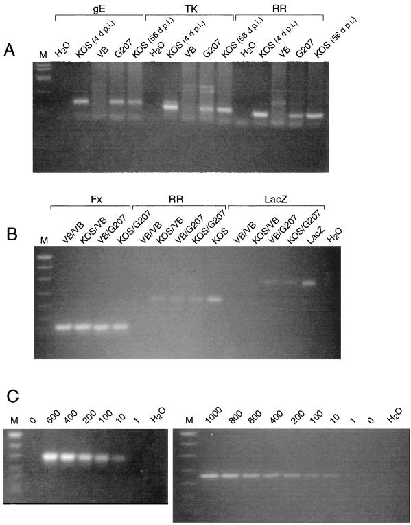
(A) Persistence of viral DNA in the mouse brain after i.c. inoculation of HSV-1. DNA was isolated from the brains of the following animals: KOS (6 × 10³ PFU) at time of death, 4 days p.i. (d p.i.) (lanes 2, 7, and 12); VB at 56 days postinoculation (lanes 3, 8, and 13); G207 (107 PFU) at 56 days p.i. (lanes 4, 9, and 14) and KOS (103 PFU) survivor at 56 days p.i. [KOS (56 d p.i.)] (lanes 5, 10, and 15). Isolated DNA was amplified with the gE (lanes 1 to 5), TK (lanes 6 to 10), and RR (lanes 11 to 15) primer pairs (Table 1), separated by agarose gel (2.5% NuSieve) electrophoresis, and visualized with ethidium bromide. The controls (lanes 1, 6, and 11) contained water in place of DNA. DNA size markers (M) are as in panel B. (B) Detection of HSV DNA sequences in the brain by PCR. DNA was isolated from the brains of animals listed in Table 4, after injections (first/second) as indicated above the lanes. Isolated DNA was amplified with the Fx (lanes 1 to 4), RR (lanes 5 to 9), and LacZ (lanes 10 to 14) primer pairs (Table 1), separated by agarose gel (2.5% NuSieve) electrophoresis, and visualized with ethidium bromide. The DNA size markers (M) are MW VIII from Boehringer Mannheim (501/489, 404, 320, 242, 190, 147, and 127 bp), the positive control for KOS (lane 9) is 100 PFU equivalents of KOS mixed with mouse brain, the positive control for LacZ (lane 14) is 10 ng of pHCL (31), and the negative control (lane 15) is water in place of DNA. The LacZ primer pair uniquely detects G207 DNA. (C) Detection sensitivity of PCR assay. HSV-1 G207 (left) or KOS (right) was mixed with mouse brain, and DNA was isolated. DNA size markers (M) are as in panel B. G207 DNA (0, 600, 400, 200, 100, 10, and 1 PFU equivalents), and water in place of DNA in lane 8, was amplified with the LacZ primer pair (Table 1) (left); KOS DNA (1,000, 800, 600, 400, 200, 100, 10, 1, and 0 PFU equivalents), and water in place of DNA in lane 10, was amplified with the RR primer pair (Table 1) (right).

