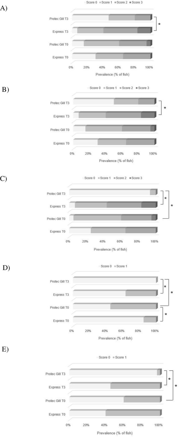
Fig 6. Gill histology.

Histopathological analysis of gill lesions analysed in Atlantic salmon gill tissue at the beginning of the study (T0) and at the end (T3): (A) epithelial and mucous hyperplasia, (B) lamellar fusion, (C) tissue degeneration/necrosis, (D) oedema, (E) pathogen load. Results are reported as prevalence percentage of fish with a specific score (from 0 to 3); n = 20 (T0), n = 30 (T3), * p < 0.05.
