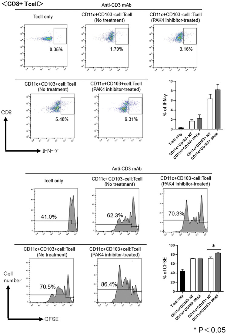
Figure 8.
T-cell stimulatory capacity of dendritic cells (DCs) following administration of PAK4 inhibitor in oral squamous cell carcinoma (OSCC) tumour-bearing mice. Splenic CD3+ T-cells were isolated from naïve C3H/HeN mice and labelled with carboxyfluorescein succinimidyl ester (CFSE). DCs (CD11c+CD103− cells and CD11c+CD103+ cells) were sorted from the tumours of OSCC tumour-bearing mice. CFSE-labelled T-cells were co-cultured with DCs in the presence of 0.1 μg/mL of an anti-CD3 Ab for 72 h at a T cell:DC ratio of 10:3. The CFSE fluorescent dye and the IFN-γ-producing T-cells among CD8+ T-cells were analysed using flow cytometry. Representative histograms and bar graphs of overall results are shown; p < 0.05, control vs PF-3758309.

