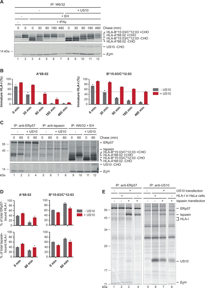
Figure 2. US10 blocks human leucocyte antigen class I (HLA-I) interaction with the peptide loading complex (PLC).
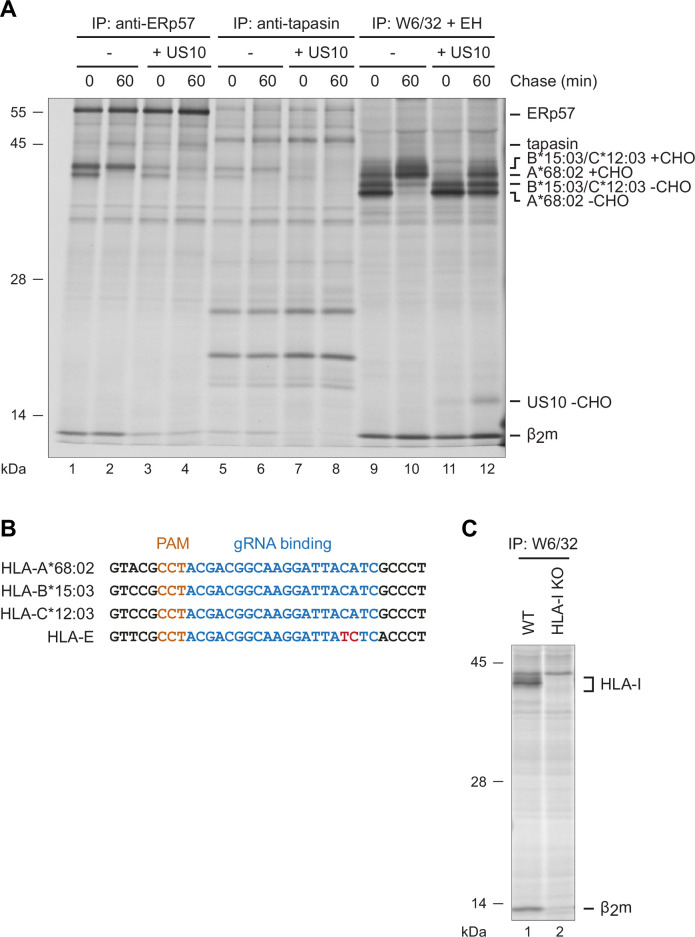
(A) Control HeLa cells or cells stably expressing US10 were induced by IFNγ overnight and subsequently metabolically labeled for 30 min and chased as indicated. After immunoprecipitation by W6/32, proteins were digested by EndoH (-CHO, deglycosylated proteins; +CHO, resistant glycans) as indicated and separated by SDS-PAGE. Labeled proteins were detected by autoradiography (Figure 2—source data 1). (B) The intensities of single HLA-I heavy chain (HC) bands in (A) were quantified, and the percentage of immature molecules compared to the total amount (sum of immature and mature) was calculated and depicted from two independent experiments (biological replicates). (C) Immunoprecipitation from HeLa cells or cells stably expressing US10 was performed as in (A) but with modified chase times and without IFNγ treatment. Antibodies applied for immunoprecipitations are indicated (Figure 2—source data 2). (D) Band intensities of HLA-I HCs in anti-ERp57 and anti-tapasin immunoprecipitations from (C) were quantified and the amount of the HLA-A*68:02 HC (left panel) and HLA-B*15:03/-C*12:03 HC (right panel) was calculated as the percentage of total PLC-bound HLA-I (sum of both HC bands). Dots represent individual values from two independent experiments (biological replicates). (E) Wild-type or HLA-I KO HeLa cells were transiently transfected with US10 and tapasin-expressing plasmids as indicated. At 20 hr post-transfection, cells were metabolically labeled for 3 hr. Immunoprecipitation was performed with anti-ERp57 or anti-US10 antibodies (Figure 2—source data 3). One of two independent experiments is shown in panels (A), (C), and (E).


