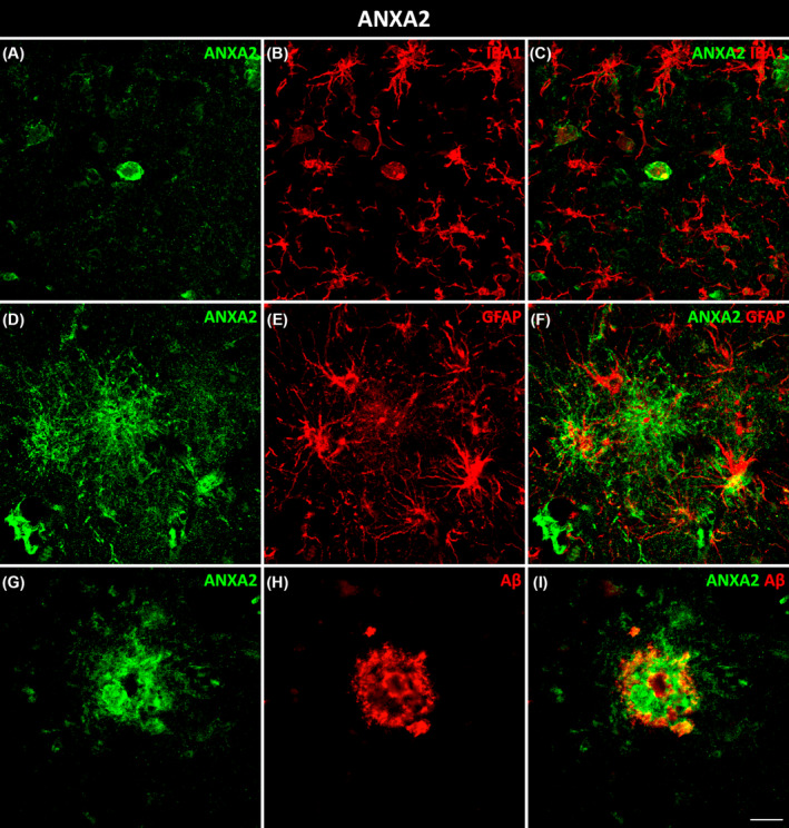
FIGURE 5.

Glial and pathological colocalization of ANXA2. Double immunofluorescence staining of coronal sections of the human EC stained for ANXA2 and IBA1 for microglia (A–C), GFAP for astrocytes (D–F), and Aβ (G–I) in AD cases. Scale bar = 20 μm. EC, entorhinal cortex.
