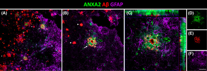
FIGURE 6.

Triple colocalization of ANXA2, Aβ plaques and astrocytes in AD. (A) Coronal section of the human EC immunofluorescently stained for ANXA2, Aβ and GFAP for astrocytes and detail (B). Arrows and arrowheads indicate Aβ plaques with and without ANXA2, respectively. Orthogonal view of the z‐stack (C) and each of its channels in green ANXA2 (D), red Aβ (E), and purple astrocytes (F). Scale bars for A = 70 μm, B = 35 μm, C = 20 μm, and D–F = 55 μm. Aβ, amyloid‐β; AD, Alzheimer's disease; EC, entorhinal cortex.
