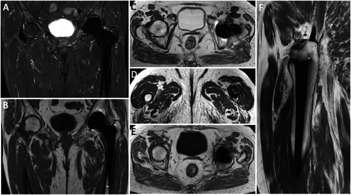
Fig. 1.
Pelvis MARS MRI of an asymptomatic 69-year-old female with normal left THA. Coronal STIR (A), coronal T1-weighted (B), axial T2-weighted (C and D), axial T1-weighted (E) and sagittal T2-weighted (F) images. No distortion or incomplete fat suppression is observed in these images. Periprosthetic bone is well-depicted without edema or osteolysis. MR images do not highlight other signs of painful THA like effusion, synovitis, collections, pericapsular edema, or masses. Further, no enlarged loco-regional lymph nodes are seen

