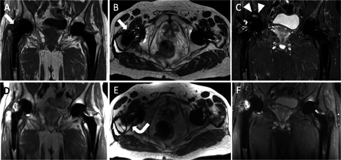
Fig. 3.
Pelvis MRI performed with MARS (A coronal T1-weighted; B axial T2-weighted; C coronal STIR) and MAVRIC (D coronal proton density-weighted; E axial T2-weighted; F coronal STIR) sequences. MAVRIC images are a bit blurred if compared with MARS, but reduce artifacts and signal loss improving imaging clarity close to metal components. Note the signal loss (arrows) near the prosthetic neck (A, B, C) and poor fat suppression in the acetabulum (arrowheads, C) on MARS images. Also, MAVRIC is more effective in depicting the right THA effusion (curved arrow, E)

