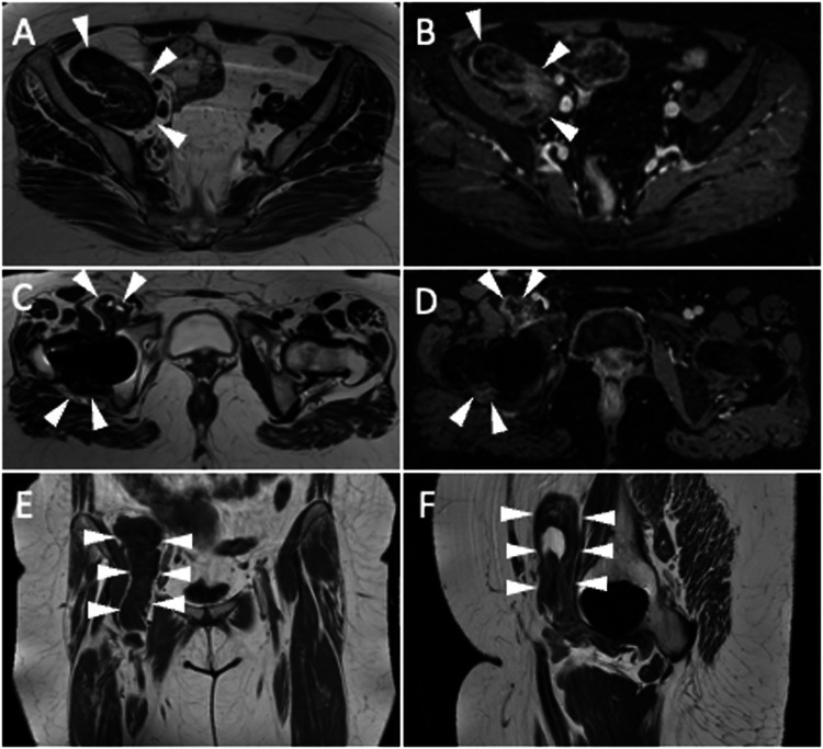
Fig. 9.
Pelvis MRI of a 58-year-old female who underwent prosthetic replacement of the right hip due to intra-articular tenosynovial giant cell tumor and experienced relapse of disease. Unenhanced axial T2-weighted (A, C), post-contrast fat-suppressed 3D GRE T1-weighted (B, D), unenhanced coronal T1-weighted (E), and unenhanced sagittal T2-weighted (F) images show periprosthetic tissue (arrowheads), located around THA neck and within the iliopsoas bursa, with T1- and T2-hypointensity and strong enhancement, similar to pseudotumor

