
Figure 2.
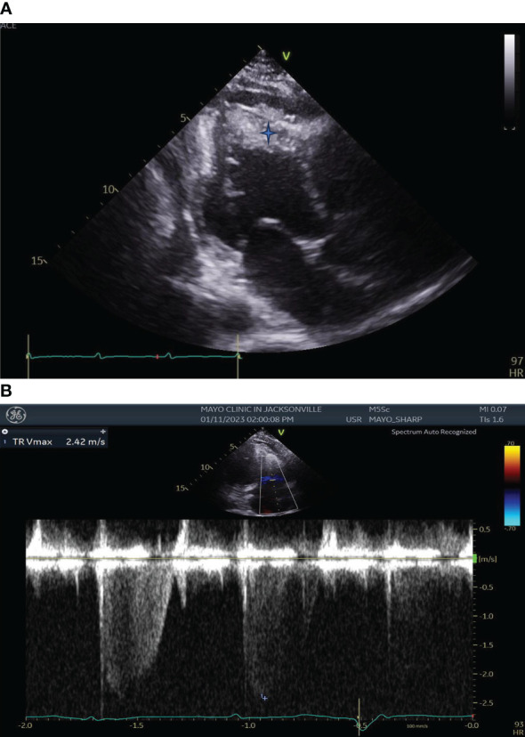
(A) Transthoracic Echocardiography showing left ventricular wall motion abnormalities, enlarged right ventricular chamber size with biventricular apical thrombi (Blue star). (B) Transthoracic Echocardiography showing severe Tricuspid Regurgitation.