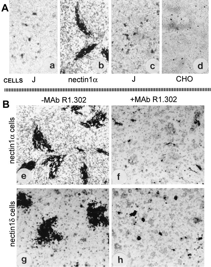
FIG. 1.
Cell-to-cell spread of HSV-1 occurs in nectin1α- or nectin1δ-expressing cells (B) but not in the receptor-negative J or CHO cells (A). (a) Micrograph of J cells transfected with HSV-1(F) DNA shows only singly infected cells or small aggregates. (b) Micrograph of stable transformants of J cells expressing nectin1α transfected with HSV-1(F) DNA shows presence of plaques. (c and d) Infectious center assay. Monolayers of nectin1α cells infected with R8102 were trypsinized and seeded onto recipient monolayers of J (c) or CHO (d) cells. Note the absence of plaques. (B) Plaque formation in nectin1α or nectin1δ cells infected with R8102 and inhibition by MAb R1.302. (e and f) Micrographs of nectin1α-expressing J cells infected with R8102, unexposed (e) or exposed to MAb R.1302 (f). (g and h) Micrographs of nectin1δ-expressing J cells infected with R8102, unexposed (g) or exposed to MAb R1.302 (h). Note that plaque size is greatly reduced in panels f and h relative to size in panel e or g. Infected cells were detected by immunostaining with polyclonal antibody to gM (a and b) or by X-Gal staining (c to h). Pictures were taken in an Axioplan Zeiss microscope. All pictures are at the same magnification.

