
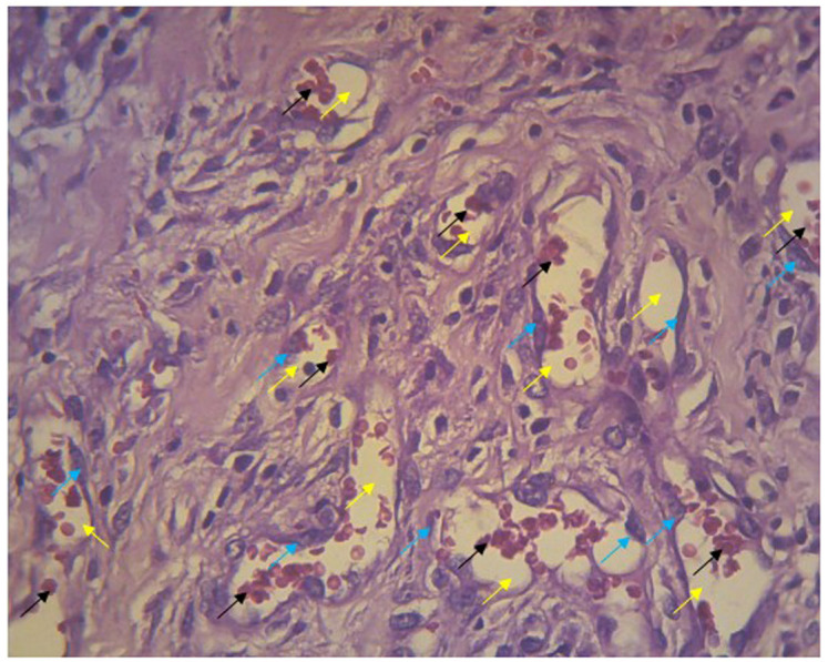
Figure 5. Blood vessels in the histologic preparation.
The walls of lumen vessels (yellow arrows) are formed by endothelial cells (blue arrows) and contain erythrocytes (black arrows). This picture was taken from experiments with gel 96%.
Official websites use .gov
A
.gov website belongs to an official
government organization in the United States.
Secure .gov websites use HTTPS
A lock (
) or https:// means you've safely
connected to the .gov website. Share sensitive
information only on official, secure websites.
The walls of lumen vessels (yellow arrows) are formed by endothelial cells (blue arrows) and contain erythrocytes (black arrows). This picture was taken from experiments with gel 96%.