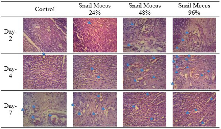
Figure 6. Angiogenesis in the wound area on days two, four, and seven, as observed from histological preparations with hematoxylin–eosin staining at 400× magnification.

The greatest number of new blood vessels formed was observed on day four following the application of 96% snail mucus gel (p = 0.000).
