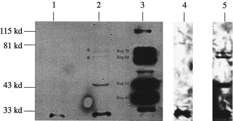
FIG. 7.
Immunoprecipitation-Western analysis of Rep expression in AAV-infected cells. HeLa cells were infected with AAV alone or Ad-AAV, and protein extracts were isolated and assayed for Rep expression using AAV Rep-specific antibody. Lanes 1, 2, 4, and 5 show Western blot analysis of AAV Rep expression from 107 HeLa cells either mock infected (lanes 1 and 4) or AAV infected (lanes 2 and 5) that were exposed for 1 min (lanes 1 and 2) or 1 h (lanes 4 and 5). Lane 3 was a positive control of 5 × 10⁶ Ad-AAV-coinfected HeLa cells demonstrating expression of all forms (78, 68, 52, and 40 kDa) of the AAV Rep proteins.

