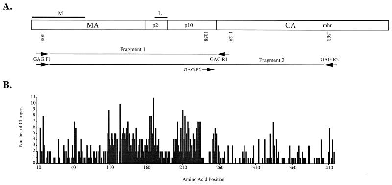
FIG. 1.
Primer locations within avian retrovirus gag genes sequenced and number of amino acid changes relative to the Gag polyprotein. Nucleotide and amino acid position numbers are relative to those of the published RSV genome (27). The known functional domains of assembly (L) and membrane binding (M) are indicated above the gag gene diagram. (A) Fragment 1 indicates the region sequenced for 102 retrovirus clones, and fragment 2 indicates the region sequenced for 40 retrovirus clones. Numbers below the diagram are nucleotide positions, starting at the 3′ base of primers. (B) Graphical representation of the number of amino acid changes over the region sequenced. Amino acid changes were determined from phylogenetic tree branch lengths as described in the text.

