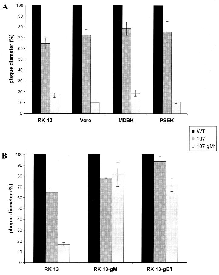
FIG. 5.
Plaque sizes of PrV-107 and PrV-107-gM−. RK13, Vero, MDBK, and PSEK cells (A), and RK13, RK13-gM, and RK13-gE/I cells (B) were infected under plaque assay conditions with PrV-107 and PrV-107-gM−. Two days after infection, plaque diameters were measured microscopically and compared with the average diameter of plaques induced by parental PrV-Be (WT, solid bars), which was set at 100%. Average values and standard deviations after measurement of at least 30 plaques in three independent experiments each are indicated.

