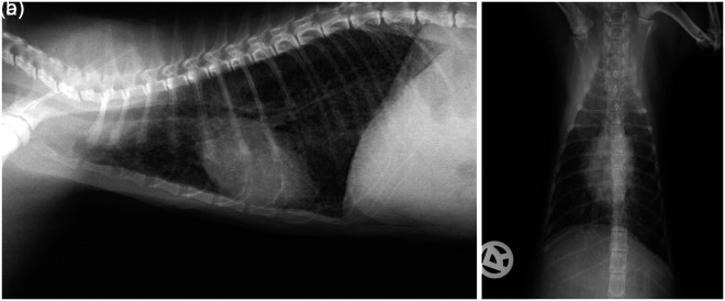
Figure 1.
Thorax, right lateral (a) and dorsoventral (b) view of cat 1 at presentation. The lungs were well inflated and the diaphragm flattened, which may represent dyspnoea. There was evidence of a generalised miliary interstitial pattern and peribronchial cuffing throughout the lungs, and a curvilinear, interrupted, mineral opacity line superimposed on the cardiac silhouette in the area of the ascending aorta and the left ventricular outflow tract, as well as evidence of bronchial mineralisation. Differential diagnosis for the generalised interstitial pattern included interstitial mineralisation secondary to hypercalcaemia, bronchopneumonia and, less likely, neoplasia secondary to a round cell tumour. Fungal pneumonia, metastatic neoplasia or uraemic pneumonitis were considered unlikely

