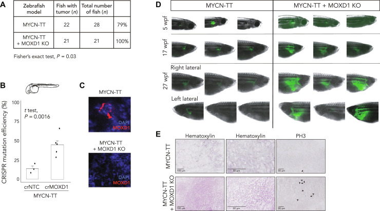
Fig. 4. MOXD1 knockout (KO) accelerates tumor penetrance in zebrafish.
(A) Summary of MYCN-TT and MYCN-TT + MOXD1 KO zebrafish with tumors. P value by Fisher’s exact test as indicated. (B) CRISPR mutation efficiency in MYCN-TT + MOXD1 KO and MYCN-TT zebrafish as determined by a tumor sample analysis on Miseq and analyzed using CRISPResso2.0 software (http://crispresso2.pinellolab.org/submission). (C) Confirmation of CRISPR-Cas9–mediated knockout of MOXD1 at protein level in tumors dissected from MYCN-TT only and MYCN-TT + MOXD1 KO zebrafish. Staining of MOXD1 by immunofluorescence. DAPI was used to counterstain the nuclei. (D) epresentative images of zebrafish at 5, 17, and 27 wpf. At week 27, each fish was photographed from both sides (right and left lateral sides). Cancer cells were visualized by fluorescence. Note that the images are not from the corresponding fish at each time point, but randomly picked from the whole population. (E) Hematoxylin staining to visualize the tissue of MYCN-TT and MYCN-TT + MOXD1 KO zebrafish tumors as well as staining with phospho-Histone H3 (PH3) visualizing mitosis. Arrowheads denote mitotic cells.

