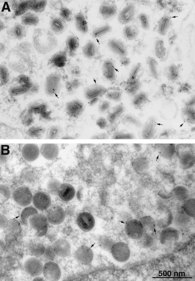
FIG. 6.
Immunogold labeling of viral membranes with antibody to the HA tag. BSC-1 cells that had been infected with vA14.5L-HA for 24 h were fixed in paraformaldehyde, cryosectioned, and incubated with a mouse MAb to the HA tag, rabbit IgG to mouse IgG, and then with 10-nm-diameter gold particles conjugated to protein A. Electron microscopic images are shown with a 500-nm marker. (A) Field containing large numbers of IEV. (B) Field containing large numbers of immature virons. Arrows point to examples of gold particles.

