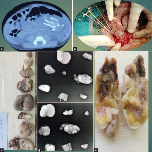
Figure 1.
(a) Contrast-enhanced computed tomography abdomen showing dense calcification or ossification seen within the cystic lesion, (b) Intraoperative picture showing fetuses being taken out, (c) Film showing all eight fetuses together, (d) Anteroposterior and lateral X-ray views of all eight fetuses, (e) Cut section of a fetus

